MRAs and MRIs are both painless and generally noninvasive tools used for diagnostic imaging. MRAs or MRIs may be used to view bones, tissues, blood vessels, arteries, or organs inside the body. As they are so closely related, it can be difficult for patients to understand the difference between an MRI and an MRA. Upon closer inspection, however, there are plenty of differences between the two you can notice despite all of the similarities. Read on to learn more about the difference between these two imaging tools and what they are used for. NeuroSkeletal Imaging Institute is a top independent imaging center with locations in Orlando, Melbourne, and Merritt Island. If you’re looking for “MRI centers near me,” call NeuroSkeletal Imaging Institute today!
How MRI Imaging Tools Work
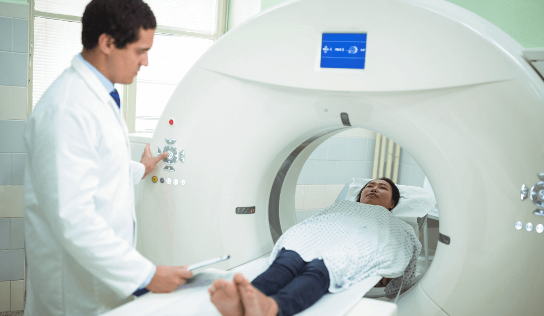
An MRI machine creates a strong magnetic field in the body, then sends signals to a computer that produces a series of these images. Every image will show a thin slice of their body. The computer then compiles these slices into a 3-D picture. Before any MRIs, the doctor will contrast dye into a vein, which allows them to more easily see inside the structure of your body. Gadolinium is the dye that doctors typically use, and may taste like metal. When taking an MRI, you lie down on a table, which slides in and out of the MRI machine. The technologists may use straps to hold you gently and help keep you still. They may have your entire body inside the machine, or just a part of it, depending on the kind of MRI you’re receiving. You may hear loud tapping, banging, thumping, or knocking noises, which the machine makes while creating the energy to take images. The MRI testing generally lasts around 20 and 90 minutes, depending on where on your body your doctor needs to scan. If you are looking for “MRI centers near me,” NeuroSkeletal Imaging Institute promises quality customer service and top-notch equipment.
How MRAs Work
MRA stands for magnetic resonance angiography, and is a medical test which assists doctors in diagnosing medical diseases and conditions of the blood vessels so that they can treat them. Radio frequency waves, a powerful magnetic field, and a computer create detailed images of your body’s major arteries during an MRA. Magnetic resonance angiography doesn’t use ionizing radiation like in x-rays. An MRA can be done with or without a contrast material. The technologist may administer the contrast material in a vein of your arm through a small intravenous (IV) catheter. Similar to an MRI, you’ll lie flat on a table that slides into a chamber, where radio waves and magnetic fields create images. Here at NeuroSkeletal Imaging Institute, an MRA takes around 30 to 90 minutes on average.
Contact Us Today
MRIs and MRAs are both imaging tools commonly used by physicians to diagnose diseases and conditions. NeuroSkeletal Imaging Institute is an independent imaging center with locations in Orlando, Melbourne, and Merritt Island. Call NeuroSkeletal Imaging Institute if you’re looking for “MRI centers near me” today!

